
All About Cerebral Vision Impairment

A side-profile photo of a young boy looking forward with a gentle smile, his chin resting on his hand. Superimposed over his head is a glowing, semi-transparent 3D illustration of a human brain and eye. A vibrant, multicolored pathway of light connects the eye to the back of the brain, visually representing the optic nerve and the process of sight.
Cerebral Visual Impairment (CVI) is the most common cause of vision impairment among children in developed countries, yet few people have heard of it and it remains extremely underdiagnosed. September is CVI Awareness Month, so we thought it was worth creating a blog post discussing all things CVI.
What is CVI?
When people think of vision impairment they tend to think of problems with the eyes, and this is understandable since this is the most obvious part of the body that we use for vision. However, eyes are the beginning part of the apparatus that we use for seeing.
Seeing begins when light enters the eyes and is focused onto the retina at the back of the eye. The retina detects this light and converts it into electrical signals, which then travel along the optic nerves towards the brain. Once these signals reach the brain, they are processed in several different areas that work together to turn them into the visual world we experience. The brain has to recognise what we are looking at, work out where things are, judge movement and distance, separate important information from background clutter, and connect what we see with our memories and other senses. In other words, the eyes collect the visual information, but it is the brain that makes sense of it. This is why it is possible for someone to have healthy eyes and still experience significant difficulties with vision.
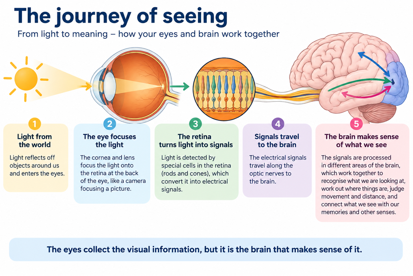
An educational infographic titled "The journey of seeing" showing how the eyes and brain work together. The diagram illustrates a five-step process from left to right:
1. Light from the sun enters a diagram of a human eye.
2. Light passes through the cornea and lens, focusing on the back of the eye.
3. A zoomed-in view shows microscopic cells in the retina (rods and cones) converting light into electrical signals.
4. Optic nerves carry these signals from the eye straight to a diagram of a human brain.
5. Colored arrows inside the brain show different areas processing the signals to make sense of the images.
A summary banner at the bottom reads: "The eyes collect the visual information, but it is the brain that makes sense of it."
A person with Ocular visual impairment has differences in their eye structure, either from birth, injury, or eye disease, which prevent light from the world being successfully focused on the retina, converted to electrical signals, and travelling through the optic nerves to the brain.
A person with Cerebral visual impairment has differences in their brain structure, either from birth, injury, or an underlying condition, which prevents the brain successfully interpreting the electrical signals it receives from the eyes.
And for some people, there can be issues with both the eyes and the brain – these people have both ocular and cerebral visual impairment.
What does CVI look like?
Parents and carers may notice atypical ‘visual’ behaviours, such as:
Difficulty finding a toy, food item or person in a busy or cluttered environment
Seeing something easily on its own, but struggling when several objects are presented together
Difficulty recognising familiar people, particularly out of context or in a crowd
Becoming overwhelmed, tired or distressed in visually busy places such as shopping centres or classrooms
Difficulty judging steps, kerbs, changes in floor level or uneven ground
Bumping into objects or people, particularly on one side
Difficulty noticing or following fast-moving objects
Taking longer than expected to look at or respond to something visually
Looking away from an object while reaching for it, rather than looking and reaching at the same time
Using peripheral vision or consistently turning the head to look at things
Finding objects easier to see when they are moving, brightly coloured or presented against a plain background
Difficulty recognising pictures, shapes or objects despite being able to see them
Losing their place when reading or scanning a page
Finding vision noticeably harder when tired, unwell, distracted or in unfamiliar surroundings
Relying heavily on touch, sound, memory or familiar routines to work out what is happening around them
No single behaviour means that a child has CVI. What is important is the overall pattern of visual behaviours, particularly when they occur consistently across everyday situations.
I think my child / the person I care for might have CVI; what should I do?
It’s essential to find a professional who is familiar with and has a good understanding of CVI. Many families approach health professionals, such as optometrists or ophthalmologists, only to be told that their child's eyes are fine. And this may absolutely be the case. However, family members are left wondering why, if eyes are fine, their child still seems totally unable to see in certain situations. If this has occurred, it is definitely worth scoping out someone with a particular interest in CVI.
How is CVI diagnosed?
Historically, obtaining a diagnosis of CVI has been difficult because there has been no universally agreed definition of exactly what CVI is, or clear criteria for how it should be diagnosed. In 2023, the National Eye Institute in the US published a proposed working definition of CVI, while the Royal College of Ophthalmologists in the UK published clinical guidance outlining key features that can be used to support a diagnosis. Together, these developments have provided a much clearer pathway for identifying and diagnosing CVI.
What does this pathway look like?
In practice, obtaining a formal diagnosis of CVI can be thought of as a two-part process: a functional vision assessment and an ophthalmologist assessment.
A functional vision assessment looks beyond eyesight alone to understand how a person uses and makes sense of vision in everyday life. When assessing for possible CVI, this has two important components:
Structured history-taking – asking carefully selected questions about how the person uses their vision in everyday situations, looking for patterns of difficulty that may not be apparent during a standard eye examination.
Clinical observation – watching how the person actually uses their vision during assessment, including how they find and recognise objects, cope with visual clutter, judge space and movement, and respond as visual tasks become more complex.
Together, these build a picture of the person’s functional vision – not simply what they can see on an eye chart, but how effectively they can use visual information in the real world.
The second part is an ophthalmologist assessment. The ophthalmologist considers the functional vision findings alongside the person’s eye health, medical and developmental history and any other relevant investigations, and can formalise the diagnosis where appropriate.
For families seeking funded supports such as the NDIS, both the functional vision assessment and the ophthalmologist assessment are usually required.. At Special Eyes, we provide the functional vision assessment, document the findings in a detailed report, and can facilitate referral to an ophthalmologist familiar with CVI. We have supported numerous families through this process, helping to bring together the functional and medical evidence needed to establish and document a CVI diagnosis.
Take-home points:
If some of the behaviours described in this article sound familiar, it is worth investigating CVI
If you have been told your child’s eyes are fine, but you still feel that they experience limitations in what and how they see, this is worth exploring further.
If you have a diagnosis of ‘Likely CVI’ we can assist in obtaining a formal diagnosis, which makes it easier to advocate for funded support and recognition.
Want to learn more?
If you are keen to find out more, please reach out to us. You can email us, or call us. Our reception is usually staffed 10am-2.30pm Mondays and Thursday and we are very happy to discuss assessment options with you and answer any questions you may have. You can find more information about our service on our website, including funding information here.